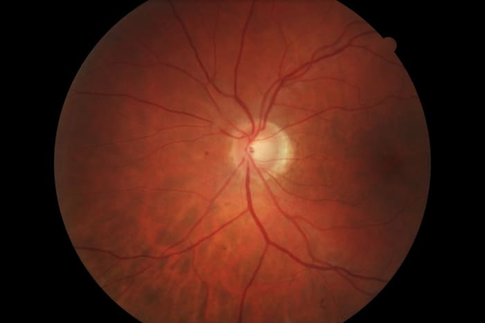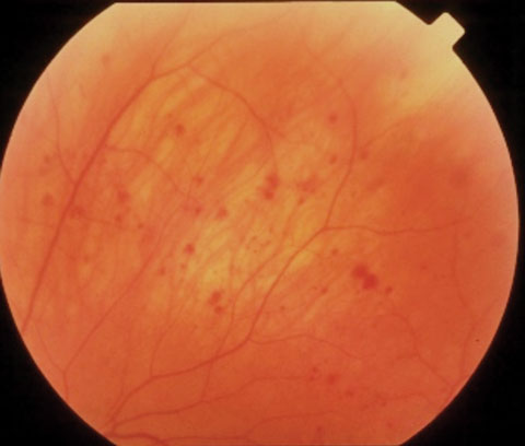

In this image, you can see a hyperreflective membrane in VR interface Sub-Segment which is exerting traction to the underneath retinal layers suggesting an Epiretinal membrane (ERM). Let’s learn to read one more similar OCT image. there are some other subtle abnormalities in this OCT image of VMT like cystic changes in the inner retina and the neurosensory detachment in deeper layers which we will discuss once we go through each of the 5 sub-segments). VR interface gives us a clue to the possible clinical diagnosis of vitreo-macular traction (VMT). Here we can easily appreciate a hyper-reflective layer in the sub-segment number 1 which is attached at fovea viz. Now let's see some examples of abnormal macular OCTs of common retinal diseases and interpret them by sub-segmenting them. Always correlate your OCT interpretation or for that matter any diagnostic imaging to your clinical examination findings. examine the patients in detail, try interpreting the clinical fundus photos before you jump in to read and interpret the OCT scans. My personal advice would be to be a good clinician first i. Henceforth in order to read and interpret a macular OCT scan rapidly, we must keep in mind the normal appearance of these 5 sub-segments.Ī thumb rule before interpreting any OCT image is to be aware of the background clinical history and fundus photo of the same eye of which you are going to interpret the OCT image. THIS IS HOW THE NORMAL MACULAR SCAN LOOKS LIKE.

Let us first review the normal macular OCT. These5 OCT sub-segments can be used for quick interpretation of macular OCT scans. And also just by looking at the diseased/damaged retinal layer, we can identify probable clinical disease. So if we segment the OCT image into 5 layer-wise sub-segments, it becomes very easy to read and interpret the retinal pathology.
#Abnormal retinal scan how to
How to quickly read a macular OCT?Īn OCT image gives you layer by layer information of retinal tissue. And so correct interpretation of OCT scans helps us in understanding the disease pathologically which in turn aids in prognosticating and making treatment decisions. In today’s practice, OCT is indisputably an essential diagnostic tool for every retinal surgeon. Optical Coherence Tomography (OCT) is a quick, non-invasive, reproducible, and optical analog of ultrasound imaging which is as good as in vivo viewing of individual retinal layers histologically.


 0 kommentar(er)
